Rationales
- NK cells are key components of innate immunity as well as adaptive immune cell therapies
- Significant limitations for NK cells from PB, CB or NK92 cell lines
- Human PSCs offer unlimited source for NK cells
- Lack of scalable, feeder-free manufacture process from PSCs to NK cells
Objective
To develop a novel manufacturing platform for the derivation of ProtoNK™ cells suitable for immune cell therapies.
Conclusions
1. We have established a 3D ProtoNK™ manufacture platform which is scalable, reproducible and efficient in generation of homogenous functional NK cells.
2. Our technology will enable mass production of ProtoNK™ cells for immuno-oncology therapies
Figure 1
Morphology of hPSC spheres (left), hemogenic endothelium (HE) rich CD31+ spheres (middle) and NK-producing spheres (right). NK cells (red arrow) were released autonomously from spheres for easy harvest. The whole process can be completed in one single-use-bioreactor at various scale.

Figure 2
>95% iPSC-derived cells are CD56+, and these CD56+ cells do not express T cell markers TCR and CD3, and B cell marker CD19, indicating iPSCderived CD56+ cells are NK cells.
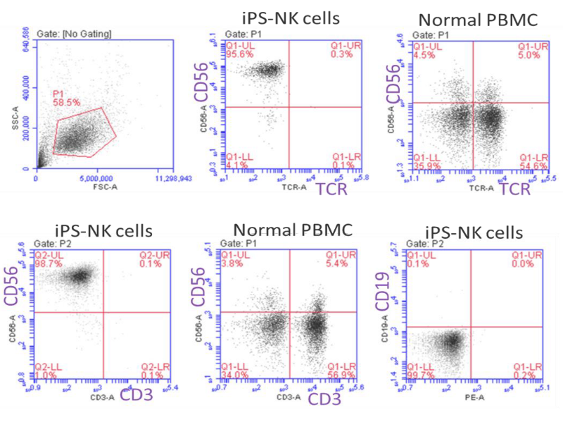
Figure 3
>95% iPS-CD56+ cells express typical NK activating receptors NKG2D, NKp44 and NKp46; and ≈30-50% of them express NK inhibitory receptors KIR2DL/DS1 and KIR2DS4; but do not express the marker of KIR3DL1/DS1; further confirming their NK cell identity.
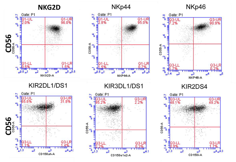
Figure 4
80% iPSC-CD56+ cells also express CD8 antigen. As CD56+CD8+ NK cells posses stronger cytotoxicity than CD56+CD8- ones, indicating iPSC-NK cells posses stronger cancer cell killing activity than regular peripheral blood NK cells.
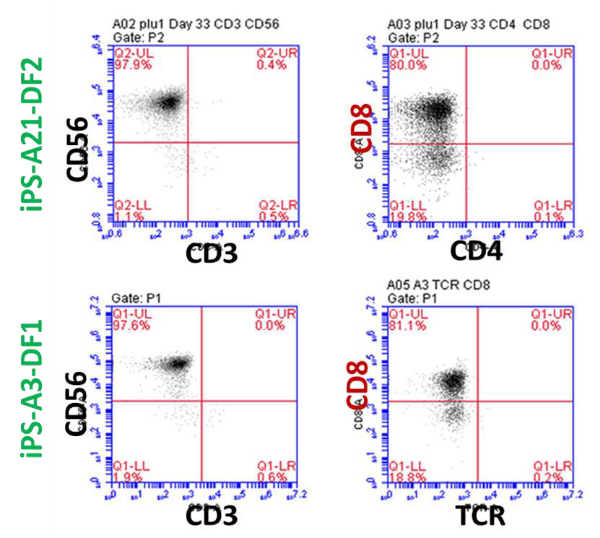
Figure 5
iPSC-NK cells were mixed with K562 cancer cells (round cells) at a ratio of 1:1. Left: healthy cell morphology at start; Right: apoptotic cell morphology at 4 hour later.
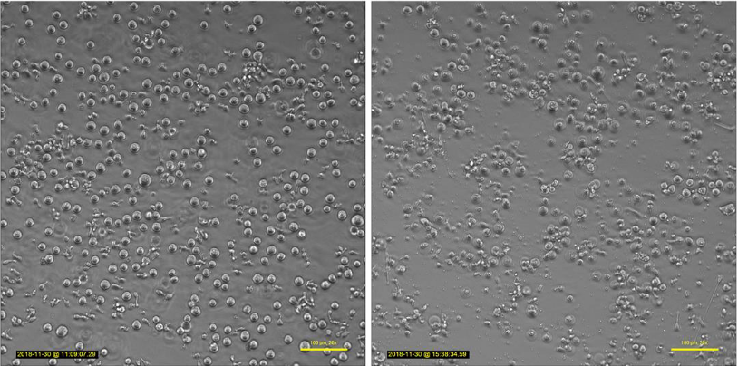
Figure 6
Top Panel: iPSC-NK cells displayed no cytotoxicity against normal human PBMC; Lower Panel: iPSC-NK showed strong cytotoxicity on K562 leukemia cells, killed >85% of them in 4 hours.
